Skip to content

NEI Research News

Thanks to the work of NEI scientists and grantees, we’re constantly learning new information about the causes and treatment of vision disorders. Get the latest updates about their work — along with other news about NEI.

Source
20 items
DNA methylation process

NEI researchers link age-related DNA modifications to susceptibility to eye disease

National Eye Institute (NEI) researchers profiling epigenomic changes in light-sensing mouse photoreceptors have a clearer picture of how age-related eye diseases may be linked to age-related changes in the regulation of gene expression.
photoreceptors

Researchers restore sight in mice by turning skin cells into light-sensing eye cells

Researchers have discovered a technique for directly reprogramming skin cells into light-sensing rod photoreceptors used for vision. The lab-made rods enabled blind mice to detect light after the cells were transplanted into the animals’ eyes.
Two boys smile at camera. Dark spot in center of image represents vision loss from AMD.

NIH Researchers Home in on Genes Linked to Age-Related Macular Degeneration

National Eye Institute scientists led a collaborative study and zeroed in on genes associated with age-related macular degeneration (AMD), a leading cause of vision loss and blindness among people age 65 and older.
Image of a mouse retina

NIH scientists deploy CRISPR to preserve photoreceptors in mice

Silencing a gene called Nrl in mice prevents the loss of cells from degenerative diseases of the retina, according to a new study. The findings could lead to novel therapies for preventing vision loss from human diseases such as retinitis pigmentosa.
Retinal degeneration in mice

Drugs already on market prevent light-induced retinal degeneration in mice

Combinations of Food and Drug Administration-approved drugs protect against the loss of cells required for vision in a mouse model of blinding retinal diseases.
Rods and Cones image

NIH Vision Scientists Test Theory of How Rods in our Retina Originated

Retinas from our earliest vertebrate ancestors had cone-like photoreceptors, presumably allowing them to see in daylight, but little ability to see at night.
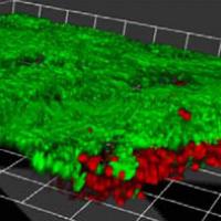
The researchers developed a technique to study live cells in patches of retina, shown here by confocal microscopy. After 2 hours, the vast majority of the cells are alive (green); only a few cells at the perimeter have died (stained red with a dye).

Retinal Cells Work with Little Reserve Energy; May Explain Vulnerability to Eye Diseases

Our eyes are especially demanding when it comes to energy: Along with our brain, they require a substantial amount of power to keep them functioning and healthy.
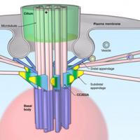
Individual cilia grow from the basal body of a cell with assistance from distal and subdistal appendages (Credit: Shobi Veleri, Ph.D., National Eye Institute)

NIH study reveals gene critical to the early development of cilia

Researchers at the National Eye Institute (NEI) have described the functions of a gene responsible for anchoring cilia — sensory hair-like extensions present on almost every cell of the body.
Two boys smile at camera. Dark spot in center of image represents vision loss from AMD.

Three studies point to same risk gene for age-related macular degeneration

Three studies reported in Nature Genetics have converged on the same gene as a rare, but powerful risk factor for age-related macular degeneration (AMD), a common cause of vision loss in older people.
National Eye Institute logo.

Additional Genes Associated with Age-Related Macular Degeneration Identified

A large genetic study of age-related macular degeneration (AMD) has identified three new genes associated with this blinding eye disease-two involved in the cholesterol pathway.