Skip to content

NEI Research News

Thanks to the work of NEI scientists and grantees, we’re constantly learning new information about the causes and treatment of vision disorders. Get the latest updates about their work — along with other news about NEI.

Source
63 items
Graphic with stylized molecules

Machine learning helps grow artificial organs

Researchers have developed a neural network capable of recognizing retinal tissues during the process of their differentiation in a dish.
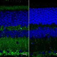
Fluorescent images of mouse retinal layers

Retinal Texture Could Provide Early Biomarker Of Alzheimer’s Disease

Biomedical engineers at Duke University have devised a new imaging device capable of measuring the various layers of the retina at the back of the eye, which could be used to detect Alzheimer's disease.
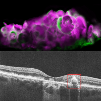
Images of dry AMD retina

NIH researchers discover tooth-enamel protein in eyes with dry AMD

A protein that normally deposits mineralized calcium in tooth enamel may also be responsible for calcium deposits in the back of the eye in people with dry age-related macular degeneration (AMD).
Grantee News

Novel technique helps explain why bright light keeps us awake

Researchers at the Salk Institute and UC San Diego discover a way to make electron microscopy more detailed and precise by visualizing the activation of brain circuits over long distances.
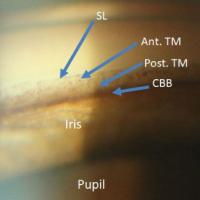
Trabecular meshwork

First accurate images of glaucoma-related eye structure taken by adapting telescope technology

Using methods originally developed by astronomers to view stars more clearly through Earth's atmosphere, optometry researchers at Indiana University have taken the first undistorted microscopic images of a part of the eye involved in glaucoma.
Grantee News

Navigating "Neuralville": Virtual town helps map brain functions

Using a virtual town, psychologists at Emory University have found that the human brain uses three distinct systems to perceive our environment.
Yellow, red and purple multi-colored mosaic

Imaging Method Reveals Long-lived Patterns in Cells of the Eye

Cells of the retinal pigment epithelium (RPE) form unique patterns that can be used to track changes in this important layer of tissue in the back of the eye, researchers at the National Eye Institute (NEI) have found.
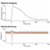
Graphs comparing neuronal signal detected by multiple methods.

Researchers design technology that sees nerve cells fire

Researchers at Stanford University, Palo Alto, California, have created a noninvasive technology that detects when nerve cells fire based on changes in shape.
Images show multimodal technique using adaptive optics and angiography to simultaneously see photoreceptors (left), retinal pigment epithelial cells (center), and choriocapillaris in the living human eye.

NIH scientists combine technologies to view the retina in unprecedented detail

By combining two imaging modalities—adaptive optics and angiography—investigators at the National Eye Institute (NEI) can see live neurons, epithelial cells, and blood vessels deep in the eye’s light-sensing retina.
Grantee News

Handheld Probe Images Photoreceptors in Children

Portable probe developed by Duke biomedical engineers makes it possible to capture images of photoreceptors in the eyes of young children.