Skip to content

NEI Research News

Thanks to the work of NEI scientists and grantees, we’re constantly learning new information about the causes and treatment of vision disorders. Get the latest updates about their work — along with other news about NEI.

Source
213 items
Grantee News

Microscopic eye movements affect how we see contrast

A team of researchers has discovered how the brain uses tiny, unconscious eye movements to pick up differences in brightness, likely by “refreshing” signals hitting the retina.
Side-by-side panels showing retinal vessels. Left panel filled with green, right panel much less green.

Faulty molecular master switch may contribute to AMD

A signaling pathway controlled by transforming growth factor β (TGF-β) could be involved in the progression of age-related macular degeneration (AMD).
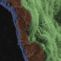
A scanning electron micrograph image shows a polarized RPE monolayer on a biodegradable scaffold. The image is colored to highlight the scaffold in blue, three RPE cells (brown), and the apical processes of cells in RPE monolayer are light green.

NIH researchers rescue photoreceptors, prevent blindness in animal models of retinal degeneration

Using a novel patient-specific stem cell-based therapy, researchers at the National Eye Institute (NEI) prevented blindness in animal models of geographic atrophy, the advanced “dry” form of age-related macular degeneration (AMD)...
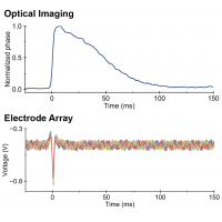
Graphs comparing neuronal signal detected by multiple methods.

Researchers design technology that sees nerve cells fire

Researchers at Stanford University, Palo Alto, California, have created a noninvasive technology that detects when nerve cells fire based on changes in shape.
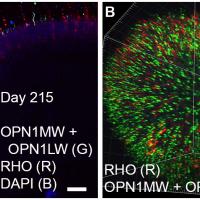
Multiphoton images show the outer segments of cone and rod photoreceptor cells in 215-day retinal organoids. Immunostaining shows a marker for cones (green) and rods (red). The “A” view shows optical sections; “B” is a reconstructed three-dimensional view.

NEI Awards Prize for Progress Toward Developing Lab-Made Retinas

The National Eye Institute (NEI) awarded $25,000 to a team led by Wei Liu, Ph.D., Albert Einstein College of Medicine, for demonstrating progress toward the development of a living model of the human retina...
Images show multimodal technique using adaptive optics and angiography to simultaneously see photoreceptors (left), retinal pigment epithelial cells (center), and choriocapillaris in the living human eye.

NIH scientists combine technologies to view the retina in unprecedented detail

By combining two imaging modalities—adaptive optics and angiography—investigators at the National Eye Institute (NEI) can see live neurons, epithelial cells, and blood vessels deep in the eye’s light-sensing retina.
NEI Audacious Goals Initiative for regenerative medicine in vision identity mark

Five research teams will develop new models for eye disease research

The National Eye Institute (NEI), part of the National Institutes of Health, has awarded grants to five multi-disciplinary teams to develop new disease models for a range of eye conditions.
Grantee News

Handheld Probe Images Photoreceptors in Children

Portable probe developed by Duke biomedical engineers makes it possible to capture images of photoreceptors in the eyes of young children.
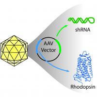
Illustration of icosahedral virus capsid, containing circular AAV vector. Arrows point from green and blue colored regions of the vector to shRNA and rhodopsin protein, respectively.

Researchers find potential new gene therapy for blinding disease

Scientists funded by the National Eye Institute (NEI) report a novel gene therapy that halts vision loss in a canine model of a blinding condition called autosomal dominant retinitis pigmentosa (adRP).
Artistic image shows Müller glia-derived rod photoreceptors.

NIH-funded researchers reverse congenital blindness in mice

Researchers funded by the National Eye Institute (NEI) have reversed congenital blindness in mice by changing supportive cells in the retina called Müller glia into rod photoreceptors.