Skip to content

NEI Research News

Thanks to the work of NEI scientists and grantees, we’re constantly learning new information about the causes and treatment of vision disorders. Get the latest updates about their work — along with other news about NEI.

Source
214 items
A microglial cell (green) extends spider-like arms to capture and consume rod photoreceptor cells (blue). Credit: Dr. Wai Wong, NEI.

In Blinding Eye Disease, Trash-Collecting Cells go Awry, Accelerate Damage

Spider-like cells inside the brain, spinal cord and eye hunt for invaders, capturing and then devouring them. These cells, called microglia, often play a beneficial role by helping to clear trash and protect the central nervous system against infection.
Grantee News

Eye’s motion detection sensors identified

Researchers at Washington University School of Medicine in St. Louis have identified a neural circuit in the retina that enable the eye to detect movement.
Grantee News

Plugging In Your Vision's Autostabilization Feature

New research, published online May 7 in the journal Neuron, describes how axons of specialized nerve cells find their way through the brain’s maze of neurons to make the right connection.
LCA is an inherited disorder that causes vision loss in childhood. It primarily affects the functioning of the retina, the light-sensitive tissue at the back of the eye, as shown here. Photo credit: National Eye Institute

NIH-funded Study Points Way Forward for Retinal Disease Gene Therapy

Gene therapy for Leber congenital amaurosis (LCA), an inherited disorder that causes vision loss starting in childhood, improved patients’ eyesight and the sensitivity of the retina within weeks of treatment.
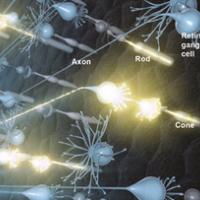
Artist's rendering of neural activity in the retina. Light that enters the eye activates rod and cone photoreceptors, which in turn activate retinal ganglion cells. Signals travel to the brain via retinal ganglion cell axons. Photo credit: National Eye Institute.

NIH Launches Research to Gaze Deeply Into Your Eyes

Five bold projects will develop new technology to noninvasively image cells of the eye in unprecedented detail.The National Eye Institute (NEI) announced the awards as part of its Audacious Goals Initiative.
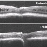
Cross-sectional images of retina from retinoschisin-deficient mice, untreated and treated with XLRS gene therapy.

NEI Human Gene Therapy Trial for Retinoschisis Underway

The National Eye Institute (NEI) recently launched the first-ever human gene therapy trial for the vision disorder X-linked retinoschisis (XLRS). Researchers are conducting the trial at the National Institutes of Health Clinical Research Center in...
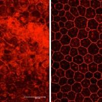
Degenerated RPE cells from mice with geographic atrophy are shown at left. Treating the mice with NRTIs protected the RPE cells. Photos courtesy of Jayakrishna Ambati, M.D.

HIV drugs show promise for “dry” AMD

A class of medications long used to curb HIV infection shows promise as a therapy for age-related macular degeneration (AMD), suggest findings from an NIH-funded study.
Grantee News

Birthday Matters for Wiring-Up the Brain’s Vision Centers

New study suggests that neurons in the developing brains of mice are guided by a simple but elegant birth order rule that allows them to find and form their proper connections.
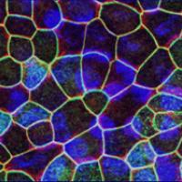
Retinal pigment epithelium derived from induced pluripotent stem cells. Photo credit: Dr. Kapil Bharti.

Gene profiling technique to accelerate stem cell therapies for eye diseases

Researchers at the National Institutes of Health have developed a technique that will speed up the production of stem-cell derived tissues.
Chichilnisky and colleagues used an electrode array to record activity from retinal ganglion cells (yellow and blue) and feed it back to them, reproducing the cells' responses to visual stimulation. Credit: E.J. Chichilnisky, Stanford.

Making artificial vision look more natural

In laboratory tests, researchers have used electrical stimulation of retinal cells to produce the same patterns of activity that occur when the retina sees a moving object.