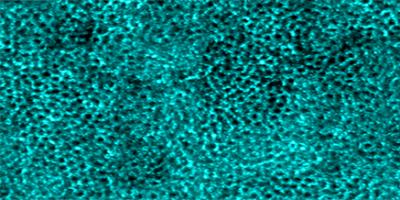
Retinal pigment epithelial cells imaged using artificial intelligence assisted adaptive optics optical coherence tomography imaging. Read the associated publication.

Erythrocytes in stasis captured within the choriocapillaris (green) and overlying parafoveal retinal capillaries (magenta). Read the associated publication.

Enlarged retinal pigment epithelial cells (left half of image) labeled with indocyanine green dye in a patient with choroideremia. Read the associated publication.

Cone photoreceptors fluorescently labeled with indocyanine green dye in a female carrier of choroideremia. Read the associated publication.
Disrupted retinal pigment epithelial cells visualized using darkfield imaging in choroideremia. Read the associated publication.

Anteriorly-displaced cone photoreceptors imaged in a patient with vitelliform macular dystrophy.

Fluorescently-labeled external limiting membrane encircling cone and rod photoreceptors, imaged using an adaptive optics translational imaging framework microscope.

Cone photoreceptor cells imaged using annular pupil illumination combined with sub-Airy disk confocal pinhole detection. Confocal reflectance (top) and non-confocal split detection (bottom) images are simultaneously acquired in a living human eye.

Widespread dark cones identified in a patient with oligocone trichromacy. Confocal reflectance image (top) showing dark cones surrounded by reflective rods and non-confocal split detection image (bottom) showing the presence of cone photoreceptors at locations where dark cones are observed.

Cone photoreceptors imaged in a human eye using adaptive optics (left) and artificially-generated (fake) images of cone photoreceptors placed in the same locations where cones are present in the left image and matched to the same size/shape, using deep learning (right). The ability to artificially-generate images can be used to efficiently create data for training deep learning algorithms.
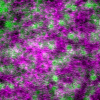
Multimodal image of retinal pigment epithelial cells. Image generated using a combination of four adaptive optics imaging modalities (magenta = summation of adaptive optics optical coherence tomography, adaptive optics infrared autofluorescence, and darkfield; green = late phase adaptive optics indocyanine green image). Individual cells labeled by indocyanine green are contained within cell outlines visualized using complementary techniques.

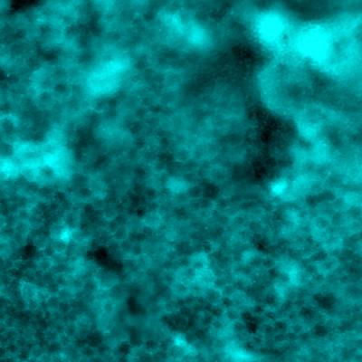
Mosaicism in the retinal pigment epithelial cells as seen by adaptive optics fluorescence microscopy. A stable fluorescence pattern is observed in human eyes following labeling by indocyanine green dye.
Read the associated publication.
View the video: Imaging method reveals long-lived patterns in cells of the eye
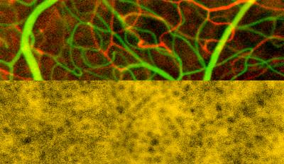
Inner retinal capillaries (top) and choriocapillaris (bottom) imaged in a living human eye using multimodal adaptive optics enhanced indocyanine green angiography.
Read the associated publication.
View the video: A look at the smallest blood vessels in the eye

Retinal pigment epithelial cells imaged noninvasively in a living human eye using adaptive optics. Image generated using a combination of multiply-scattered light (red) and intrinsic near-infrared autofluorescence (green).

Cone photoreceptor neurons (red) and retinal pigment epithelial cells labeled using indocyanine green dye (green), imaged in a living human eye using adaptive optics.

A custom-built adaptive optics ophthalmoscope, located within the Eye Clinic of the NIH Clinical Center, was used to obtain all of the images in this image gallery. For more information about the different imaging modalities contained within this instrument, click.