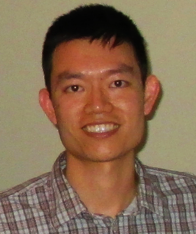
Senior Investigator
Research interests: Clinical and translational imaging
Contact Information:
Email 301-435-7821
Building 10, Room 9N240
10 Center Drive
Bethesda,
Maryland
20892-1860
Biography
Dr. Tam received a bachelor’s degree in bioengineering from the University of California, San Diego, followed by a Ph.D. in bioengineering from a joint program administered by the University of California, San Francisco and the University of California, Berkeley. His graduate studies, under the mentorship of Austin Roorda, focused on developing noninvasive microvascular imaging tools to study diabetic retinopathy. He then went on to pursue postdoctoral training in the lab of Melike Lakadamyali, working on stochastic optical reconstruction microscopy (STORM), a superresolution technique. Dr. Tam was recently named a Stadtman Investigator, where he and his staff are working on clinical applications of adaptive optics. He is currently a Senior Investigator.
Current research
The goal of our research is to understand the onset and progression of retinal diseases at the cellular level, using advanced optical imaging techniques such as adaptive optics. Adaptive optics is a technology for measuring and correcting the optical imperfections utilized in astronomy, microscopy, and vision science. When combined with a state-of-the-art ophthalmic imaging platform, highly detailed images of the cells in the human retina can be acquired.
The approach is to visualize healthy and diseased cells directly inside patients’ eyes to determine the sequence and timing of all the cumulative microscopic changes that give rise to clinically-significant disease phenotypes. Our research spans the development, implementation, and application of advanced optical instrumentation, as well as the acquisition, processing, and analysis of rich imaging datasets. We are particularly interested in studying the outer retina, consisting of photoreceptor neurons, retinal pigment epithelial cells, and choriocapillaris blood vessels. This multi-layered complex is not only critical for the phenomenon of vision, but also, is a useful system for modeling the in vivo interactions of neurons, epithelial cells, and vasculature within the central nervous system, in health, aging, and disease.
Selected publications
- Aguilera N, Liu T, Bower AJ, Li J, Abouassali S, Lu R, Giannini J, Pfau M, Bender C, Smelkinson MG, Naik A, Guan B, Schwartz O, Volkov A, Dubra A, Liu Z, Hammer DX, Maric D, Fariss R, Hufnagel RB, Jeffrey BG, Brooks BP, Zein WM, Huryn LA, Tam J. Widespread subclinical cellular changes revealed across a neural-epithelial-vascular complex in choroideremia using adaptive optics.(external link) Commun Biol. 2022;5(1):893.
- Li J, Wang D, Pottenburgh J, Bower AJ, Asanad S, Lai EW, Simon C, Im L, Huryn LA, Tao Y, Tam J, Saeedi OJ. Visualization of erythrocyte stasis in the living human eye in health and disease.(external link) iScience. 2023;26(1):105755.
- Das V, Zhang F, Bower AJ, Li J, Liu T, Aguilera N, Alvisio B, Liu Z, Hammer DX, Tam J. Revealing speckle obscured living human retinal cells with artificial intelligence assisted adaptive optics optical coherence tomography.(external link) Commun Med (Lond). 2024;4(1):68.
- Jung H, Liu J, Liu T, George A, Smelkinson MG, Cohen S, Sharma R, Schwartz O, Maminishkis A, Bharti K, Cukras C, Huryn LA, Brooks BP, Fariss R, Tam J. Longitudinal adaptive optics fluorescence microscopy reveals cellular mosaicism in patients.(external link) JCI Insight. 2019;4(6).
- Lu R, Aguilera N, Liu T, Liu J, Giannini JP, Li J, Bower AJ, Dubra A, Tam J. In-vivo sub-diffraction adaptive optics imaging of photoreceptors in the human eye with annular pupil illumination and sub-Airy detection.(external link) Optica. 2021;8(3):333-343.