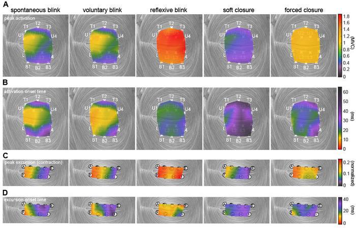
Muscle activation and movement patterns over time across the upper and lower eyelids, shown under different actions. Credit: Anatomical Engineering Group/UCLA
A blink of an eye seems natural and instantaneous, but is it? Without a functioning eyelid, the eye can become dry, irritated and eventually lose the ability to see clearly.
New research funded by the National Eye Institute at the University of California, Los Angeles has uncovered details about the muscle that controls blinking, offering a pathway toward developing blink-assisting prostheses.
Published in the Proceedings of the National Academy of Sciences, the study found that the orbicularis oculi — the muscle that controls eyelid movement — contracts in complex patterns that vary by action and move the eyelid in more than just a simple up-and-down motion.
The researchers studied how this muscle behaves differently across various actions including spontaneous blinks, protective rapid closures and squeezed shut-eye motions.
“The eyelid’s motion is both more complex and more precisely controlled by the nervous system than previously understood,” said study corresponding author Tyler Clites, Ph.D., an assistant professor of mechanical and aerospace engineering at the UCLA Samueli School of Engineering. “Different parts of the muscle activate in carefully timed sequences depending on what the eye is doing. This level of muscle control has never been recorded in the human eyelid. Now that we have this information in rich detail, we can move forward in designing neuroprostheses that help restore natural eyelid function.”
In experiments with volunteers, the researchers looked at five different ways the eyes close:
- Spontaneous blink: An automatic, unconscious blink that occurs regularly to keep the eye lubricated
- Voluntary blink: An intentional blink, as when someone is asked to blink on command
- Reflexive blink: A rapid, involuntary blink triggered to protect the eye from a collision
- Soft closure: A gentle, slow eyelid descent, similar to the beginning of sleep
- A forced closure: A deliberate squeezing of the eyelids tightly shut
To record activity in the orbicularis oculi with high precision, an ophthalmic surgeon inserted tiny wire electrodes into the eyelid. The researchers then used a motion-capture system to track eyelid movement in ultraslow motion. These tools allowed the team to measure subtle differences in eyelid movement, including speed, direction, and which part of the muscle initiated the action.
To read more, visit UCLA Samueli School of Engineering