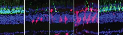
Sequence of five images, spanning 28 days, shows how regeneration happens in the zebrafish retina. Rods are shown in green, regenerating cells are shown in red, and all other cells are labeled with blue. As the rods die, regenerating cells increase and replace the lost rods.
Researchers at Vanderbilt University in Nashville, Tennessee, have discovered that in zebrafish, decreased levels of the neurotransmitter gamma-aminobutyric acid (GABA) cue the retina, the light-sensing tissue in the back of the eye, to produce stem cells. The finding sheds light on how the zebrafish regenerates its retina after injury and informs efforts to restore vision in people who are blind. The research was funded by the National Eye Institute (NEI) and appears online today in Stem Cell Reports. NEI is part of the National Institutes of Health.
“This work opens up new ideas for therapies for blinding diseases and has implications for the broader field of regenerative medicine,” said Tom Greenwell, Ph.D., NEI program officer for retinal neuroscience.
For years, vision scientists have studied zebrafish to understand their retinal regenerative capacity. Zebrafish easily recover from retinal injuries that would permanently blind a person. Early studies in zebrafish led to the idea that dying retinal cells release signals that trigger support cells in the retinal called Muller glia to dedifferentiate—return to a stem-like state—and proliferate.
However, recent studies in the mouse brain and pancreas suggest GABA, a well-characterized neurotransmitter, might also play an important role in regeneration distinct from its role in communicating local signals from one neuron to the next. Scientists studying a part of the brain called the hippocampus found that GABA levels regulate the activity of neural stem cells. When GABA levels are high, the stem cells stay quiet, and if GABA levels decrease, the stem cells start to divide, explained James Patton, Ph.D., Stevenson Professor of Biological Sciences at Vanderbilt and senior author of the new study in zebrafish retina. A similar phenomenon was reported in the mouse pancreas.
Based on these findings, Patton and his student Mahesh Rao hypothesized that GABA might be involved in the zebrafish retina’s regeneration response. To test their idea, Patton and Rao injected GABA inhibitors into undamaged zebrafish eyes and found that the fish developed a regenerative response; that is, Muller glia in the retina dedifferentiated and proliferated. Conversely, increasing GABA levels after inducing retinal damage suppressed proliferation of dedifferentiated Muller glia.
These findings supported the researchers’ hypothesis that decreased GABA signaling is a cue for regeneration in the zebrafish retina. “This is the first report to show a regenerative role for GABA in the zebrafish retina,” said Patton.
Patton and co-authors are conducting ongoing work to determine if the dedifferentiated Muller glia can turn into functional retinal cells, such as the light-activated photoreceptors. They are also exploring whether altering GABA signaling might coax a regenerative response in the retina of other species such as mice.
Patton is currently funded by the NEI Audacious Goals Initiative, a sustained effort to develop regenerative therapies for blinding diseases of the retina. The research was funded by National Eye Institute grants R01 EY024354, R21 EY019759 and P30-EY008126.
Reference: Rao, MB, Didiano D, Patton JG. 2017. Neurotransmitter-regulated Regeneration in the Zebrafish Retina. Stem Cell Reports. Pubmed