
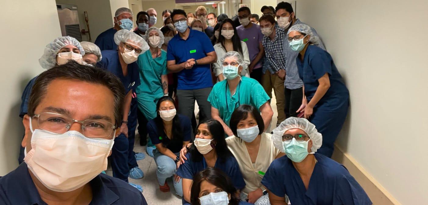
Dr. Kapil Bharti and his multidisciplinary research team in 2021, celebrating the first surgical implantation of RPE cells in their human clinical study.
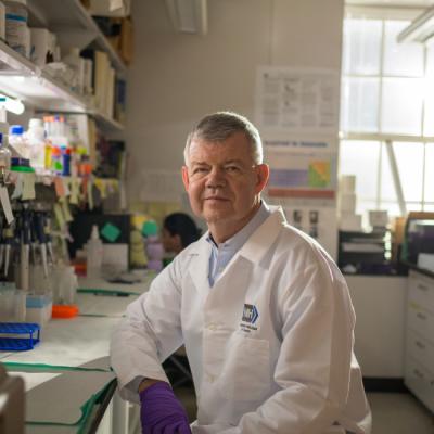
Dr. Kapil Bharti did not set out to become a vision researcher. Trained as a molecular biologist in Germany, his early career focused almost entirely on test tube-based experiments. He joined the National Institutes of Health (NIH) for postdoctoral training with the goal of working more directly with the living systems that impact everyday life.
That opportunity came in a developmental biology lab at NIH. There, researchers were studying how the body creates the retinal pigment epithelium (RPE), a thin layer of cells in the eye that is essential for sight.
“I was more fascinated by how the eye forms and how it's made.”
— Dr. Kapil Bharti
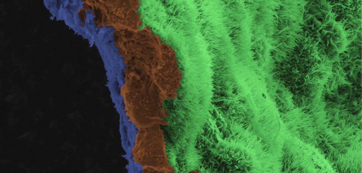
Lab-grown retinal pigment epithelial (RPE) cells are carefully arranged on a biodegradable scaffold designed to replace damaged tissue and support vision-restoring cell therapy.
How Light Becomes Sight
Vision depends on an intricate partnership among the different parts of the eye, each playing a role in turning light into sight. While photoreceptors are the light sensing cells on the retina, the RPE is the foundation for the entire process. This single layer of cells delivers nutrients from blood, removes waste, protects the photoreceptors, and regenerates their visual pigment.
That tireless workload can eventually lead to RPE breakdown, resulting in conditions like age-related macular degeneration (AMD). AMD is the leading cause of vision loss in the United States, affecting millions of Americans. In AMD, the RPE cells are often the first to fail, and they are unable to regenerate. Once they are gone, photoreceptors lose their support, and vision loss follows.
Replacing What Time Can Erode
Rather than attempting to reverse the damage, Dr. Bharti and his colleagues asked a different question: If RPE cells are the first to fail, could replacing them prevent the resulting loss of sight?
The answer began with a scientific breakthrough that seemed straight out of science fiction: induced pluripotent stem cells. Researchers create these cells in the lab from a small sample of a patient’s blood or skin, and they can then be changed to become many different tissues of the body. In Dr. Bharti’s lab, years of molecular biology and eye‑development research led to a reliable method for turning those stem cells into fully mature and functional RPE cells.
But delivering those cells safely to the eye presented a new challenge, and another opportunity for collaboration.
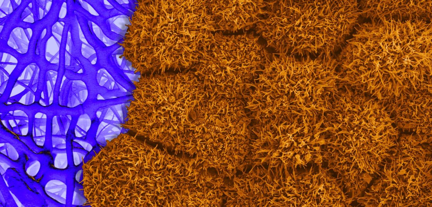
The scaffolding provides the structure that allows the cells to form the new portion of the RPE before dissolving and leaving the cells intact.
More than Meets the Eye
RPE cells function as a connected sheet bound together. Injecting new cells would destroy the structure they depend on.
To solve this, Dr. Bharti and the team turned to engineers and biomaterials experts from the nearby National Institute of Standards and Technology (NIST) to collaborators across California, London, Japan, and South Africa.
Together, they designed a paper‑thin, biodegradable scaffold made from a material already familiar to the U.S. Food and Drug Administration (FDA). The scaffold supports the cells during surgery, allowing nutrients to pass through, and then dissolves. The result is living tissue, derived from the patient’s own cells, that integrates naturally into the eye.
“There were many parallel paths that all had to come together.”
— Dr. Kapil Bharti
This work was made possible by the unique environment at the National Eye Institute (NEI). Basic scientists, stem cell biologists, engineers, regulatory experts, and clinicians work steps apart, enabling discoveries to move efficiently from laboratory to clinic. Federal investment allows researchers to pursue transformative ideas without pressure for immediate profit, laying the groundwork for even more breakthroughs to come.
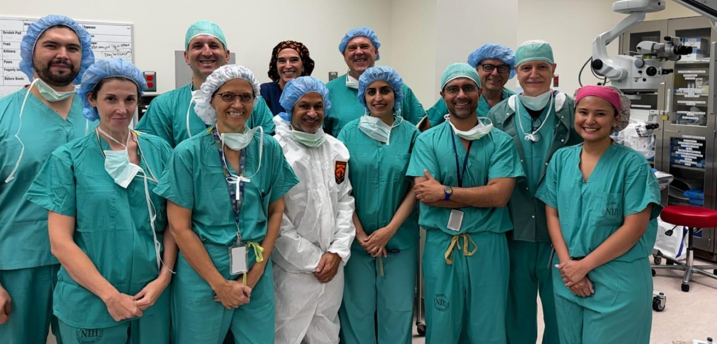
The surgical team celebrates another successful procedure as the clinical trial advances, reflecting the close collaboration between scientists, surgeons, and clinical staff working to develop new treatments for age-related macular degeneration.
Looking Ahead
Today, Dr. Bharti and the NEI team is testing RPE transplantation for advanced AMD. In the future, similar approaches may help restore sight for people living with rare inherited retinal diseases and conditions linked to severe nearsightedness.
The tools supporting this work continue to evolve, including artificial intelligence methods that help ensure cell quality and consistency.
But the core lesson remains. Progress in vision research is not the result of a single discovery. Rather, lasting impact in vision research is built on shared expertise, sustained public investment, and trust.
Vision Research in Action
A Legacy of Innovation
The first FDA-approved gene therapy, Luxturna was developed based on intramural research led by NEI scientists.