Ante la falta de fondos del gobierno federal, no se actualizará este sitio web y la organización no responderá a transacciones ni consultas hasta que se aprueben los fondos. El Centro Clínico de los Institutos Nacionales de la Salud (el hospital de investigación) permanecerá abierto. Consulte cc.nih.gov (en inglés). Infórmese sobre el funcionamiento del gobierno federal y el reinicio de las actividades en OPM.gov
Thanks to the work of NEI scientists and grantees, we’re constantly learning new information about the causes and treatment of vision disorders. Get the latest updates about their work — along with other news about NEI.
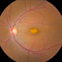
Using a new imaging technique, researchers from the National Eye Institute have determined that retinal lesions from vitelliform macular dystrophy (VMD) vary by gene mutation.
Black patients have a dramatically higher risk of advanced vision loss after a new diagnosis of primary open angle glaucoma (POAG) when compared to white patients, according to a new study from New York Eye and Ear Infirmary of Mount Sinai (NYEE).
Loss of the protein pigment epithelium-derived factor (PEDF), which protects retinal support cells, may drive age-related changes in the retina, according to a new study in mice from the National Eye Institute.
Clinical trial results from the DRCR Retina Network suggest that a specific step strategy gives results similar to starting off with the higher-priced drug.
Scientists at LSU Health New Orleans Neuroscience Center of Excellence have developed a new, experimental human cell line from retinal pigment epithelial cells.
New research from the Max Planck Florida Institute for Neuroscience demonstrates that inhibitory and excitatory neuronal circuits of the visual system develop through different processes, even if the organization of the mature circuit is similar.
In a new research study from the Max Planck Florida Institute for Neuroscience, scientists have now discovered the neuronal changes that occur during learning to improve discrimination of closely related visual images.
The eye’s light-sensing retina taps different circuits depending on whether it is generating image-forming vision or carrying out a non-vision function such as regulating pupil size or sleep/wake cycles, according to a new mouse study.
A new study from Brown University demonstrated that some regions of the cerebral cortex involved in cognitive processing and mood show sensitivity for light intensity.