
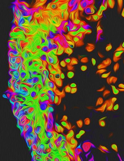
Artistic rendition of 3-dimensional retinal organoid exposed to oxidative stress demonstrating marked cell death (apoptosis; green) within retinal photoreceptors (pink). Image credit: Isabella Sodhi, McDonogh School.
Wilmer Eye Institute researchers at Johns Hopkins Medicine have found how a molecular pathway — involving oxidative stress, or an imbalance of molecular oxygen in cells, and the protein HIF-1 — contributes to what kind of age-related macular degeneration (AMD) a patient could develop.
Researchers started by focusing on oxidative stress, which can increase in the body through common factors such as aging, exposure to cigarette smoke and high-fat/high-sugar diets. Both oxidative stress and HIF-1 have been previously implicated in the development of AMD.
Specifically, investigators looked at changes in HIF-1 levels caused by oxidative stress in two eye cell populations: retinal pigment epithelium, which protects the retina and filters light, and retinal photoreceptors, nerve cells that convert light to brain signals.
When the researchers induced oxidative stress in human and rodent photoreceptors, they saw an increase in HIF-1 production, similar to their experiments in retinal pigment epithelium. However, they observed that while retinal pigment epithelial cells did not die, the photoreceptors were remarkably sensitive to oxidative stress, which caused cell death in the tissue, thus mimicking dry AMD.