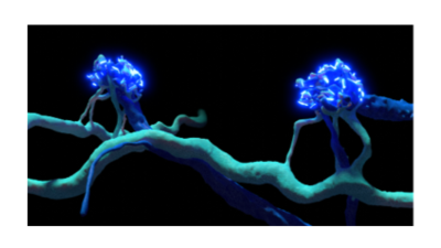
3D reconstructed neurites from S-cone selective neurons reach out to contact the only two S-cones among a field of photoreceptors. Credit: John Ball
National Institutes of Health (NIH) researchers have constructed a 3D map of neural circuitry of S-cone photoreceptors, a type of cell in our eye important for perceiving blue light. The findings provide a foothold for future studies not only on color perception, but also for studying myopia and mood disorders related to light. The work published December 1 in the journal, Proceedings of the National Academy of Sciences.
The report characterizing the S-cone circuitry is an example of a neuroscience field called connectomics, which uses a combination of high-resolution imaging, artificial intelligence, and at times, functional recording to study how the brain’s massively complex neural circuitry structure relates to its function.
“The retina is an ideal application for connectomics because it is an accessible part of the brain and because we already have a century of research into its circuits,” said the study’s lead investigator, John Ball, Ph.D., staff scientist in the Retinal Neurophysiology Section at NIH’s National Eye Institute (NEI).
S-cones are a type of photoreceptor within the light-sensitive retinal tissue at the back of the eye. Photoreceptors (cones and rods) in the retina convert light into electrical signals, which are sent to visual centers in the brain. Rods enable vision in low light conditions. Cones enable color vision, which in humans involves three cone types sensitive to wavelengths of light perceived as red, green, and blue. S-cones are well suited for detecting short wavelength light, which is perceived as blue.
In addition to helping us perceive the sky as blue, evidence increasingly suggests that S-cone photoreceptors contribute to a host of non-image forming vision. Circadian rhythms, for example, which affect sleep and mood are influenced by blue light. Blue light may also mediate eye growth during childhood and teenage years. Childhood onset myopia or near sightedness is caused when the eye grows too long from front to back causing images to form at a point in front of the retina.
“From an evolutionary perspective, many aspects of the S-cone, its circuitry and function appear to be well conserved across mammalian species, suggesting its importance for survival,” said Ball. “Yet despite being vital for survival, S-cones and their circuitry are difficult to study in part because they are relatively scarce within the retinas of the animal models we use,” he said.
Mouse models are less helpful for studying S-cones as their retinas are comprised mostly of rod photoreceptors due to their nocturnal lifestyle. For that reason, Ball and his colleagues relied on the 13-lined ground squirrel, a diurnal model with a cone-dominant retina.
Their connectomics data reconstructed the S-cone as well as its downstream points of interaction (synaptic terminals) with surrounding retinal cells. The researchers also used electrophysiology and electroretinography to detect the signaling activity of these S-cone circuits.
Future research is needed to investigate the rest of the circuitry involved in color vision in mammals with cone-dominant retinas and how a better understanding of the S-cone circuitry may lead to treatments for myopia and some mood disorders.
The work was funded by an NEI Intramural Research Program.
Reference: Zhang Y, Chen S, Qian H, Li W, and Ball JM. “S-cone specific circuitry in the outer plexiform layer of a cone-dominant mammal” published December 1, 2025 PNAS