Skip to content

Because of a lapse in government funding, the information on this website may not be up to date, transactions submitted via the website may not be processed, and the agency may not be able to respond to inquiries until appropriations are enacted.
The NIH Clinical Center (the research hospital of NIH) is open. For more details about its operating status, please visit cc.nih.gov.
Updates regarding government operating status and resumption of normal operations can be found at OPM.gov.

NEI Research News

Thanks to the work of NEI scientists and grantees, we’re constantly learning new information about the causes and treatment of vision disorders. Get the latest updates about their work — along with other news about NEI.

Source
294 items
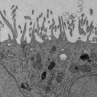
Grey scale electron microscopy image of human retinal cells.

New genetic links found to rare eye disease, opening the door to better diagnostics and potential treatments

NEI-funded research at Scripps Research Institute has turned up more than a dozen gene variants linked to MacTel, a rare eye disease. The variants are likely causing the condition to develop and worsen for a significant share of patients.
Rod and cone photoreceptors are shown in greyscale images.

NIH-led team sets new bar in retinal imaging

A team led by scientists at the National Eye Institute (NEI) has noninvasively visualized the light-sensing cells in the back of the eye, known as photoreceptors, in greater detail than ever before.

Researchers use patients’ cells to test gene therapy for rare eye disease

Scientists at the National Eye Institute (NEI) have developed a promising gene therapy strategy for a rare disease that causes severe vision loss in childhood.
Two boys smile at the camera. Asymmetric dark spots block parts of the image, representing vision loss from diabetic retinopathy.

AI algorithms detect diabetic eye disease inconsistently

Researchers from UW Medicine have found that artificial intelligence-based screening algorithms to diagnose diabetic retinopathy do not perform as well as they claim.
Dr. Emily Chew of the National Eye Institute examines a patient’s eyes. Photo credit: National Eye Institute.

Routine eye scans may give clues to cognitive decline in diabetes

Scientists at Joslin Diabetes Center now have shown that routine eye imaging can identify changes in the retina that may be associated with cognitive disorders in older people with type 1 diabetes.
Fundus photo showing medium drusen

VisionQuest Gains FDA 510(k) Clearance for the Image Quality Analyzer Software

The IQA is a software system intended for use in importing, displaying, analyzing and managing images acquired with digital fundus cameras. The patented software detects the most common causes of retinal imaging artifacts.
Image shows eye globe, optic nerve, lens, cornea, pupil, meshwork, angle, and where fluid forms and exits the front of the eye.

Cataract surgery in infancy increases glaucoma risk

Children who undergo cataract surgery as infants have a 22% risk of glaucoma 10 years later, whether or not they receive an intraocular lens implant. The findings come from the National Eye Institute (NEI)-funded Infant Aphakic Treatment Study.

Surgical and drug treatment options lead to similar outcomes for diabetic eye disease

Surgical and injectable drug approaches are equally effective for treatment of bleeding inside the eye from proliferative diabetic retinopathy (PDR), according to a National Eye Institute (NEI)-supported clinical study from the DRCR Retina Network .
3D printed model of Sars-CoV-2

COVID-19 Found in The Cornea: Are Transplants a Transmission Risk?

A multi-institutional study finds that COVID-19 can be found in post-mortem corneal tissue, highlighting the importance of the donor screening process.
Stem cell illustration

New stem cell technique offers hope for those with corneal damage

Harvard Medical School surgeons at Massachusetts Eye and Ear have replaced the ocular surface of four patients who each experienced chemical burns to one eye.