Skip to content

NEI Research News

Thanks to the work of NEI scientists and grantees, we’re constantly learning new information about the causes and treatment of vision disorders. Get the latest updates about their work — along with other news about NEI.

Source
65 items
Images show multimodal technique using adaptive optics and angiography to simultaneously see photoreceptors (left), retinal pigment epithelial cells (center), and choriocapillaris in the living human eye.

NIH scientists combine technologies to view the retina in unprecedented detail

By combining two imaging modalities—adaptive optics and angiography—investigators at the National Eye Institute (NEI) can see live neurons, epithelial cells, and blood vessels deep in the eye’s light-sensing retina.
Grantee News

Handheld Probe Images Photoreceptors in Children

Portable probe developed by Duke biomedical engineers makes it possible to capture images of photoreceptors in the eyes of young children.
Grantee News

New imaging technique by IU researchers could improve the diagnosis, treatment of glaucoma

New ophthalmoscopy method obtains high-definition, high-contrast images of translucent cells where the disease originates.
Grantee News

How the brain recognizes familiar faces

Researchers at The Rockefeller University have begun to unravel the mystery of how the brain recognizes familiar faces. Their results are published in the journal Science.
Grantee News

New Method to Detect Ultrasound with Light

A tiny, transparent device that can fit into a contact lens has a bright future, potentially helping a range of scientific endeavors from biomedicine to geology.
Grantee News

Handheld Device Takes High-Resolution Images of Children's Retinas

Engineers and physicians at Duke University have developed a handheld device capable of capturing images of a retina with cellular resolution.
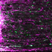
Regenerating mouse retinal ganglion cell axons (magenta and green) extending from site of optic nerve injury (left). Photo courtesy of Andrew D. Huberman.

Use it or Lose it: Visual Activity Regenerates Neural Connections Between Eye and Brain

A study in mice funded by the National Institutes of Health (NIH) shows for the first time that high-contrast visual stimulation can help damaged retinal neurons regrow optic nerve fibers, otherwise known as retinal ganglion cell axons.
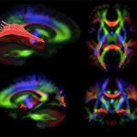
Images of the brain generated by diffusion tensor imaging, a type of MRI. The red highlights show visual pathways in the brain that deteriorate in patients with LCA, but appear to improve with gene therapy to the retina. Credit: Dr. Manzar Ashtari, University of Pennsylvania.

With LCA Gene Therapy, a Rare Glimpse of the Adult Brain Adapting to New Experience

When people lose the ability to see, how do the visual parts of the brain change in response? And if they regain their sight, are the changes reversed?
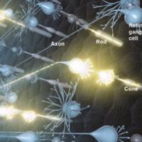
Artist's rendering of neural activity in the retina. Light that enters the eye activates rod and cone photoreceptors, which in turn activate retinal ganglion cells. Signals travel to the brain via retinal ganglion cell axons. Photo credit: National Eye Institute.

NIH Launches Research to Gaze Deeply Into Your Eyes

Five bold projects will develop new technology to noninvasively image cells of the eye in unprecedented detail.The National Eye Institute (NEI) announced the awards as part of its Audacious Goals Initiative.
Grantee News

IU School of Optometry study: New technique detects microscopic diabetes-related eye damage

Indiana University researchers have detected new early-warning signs of the potential loss of sight associated with diabetes. This discovery could have far-reaching implications for the diagnosis and treatment of diabetic retinopathy