Skip to content

NEI Research News

Thanks to the work of NEI scientists and grantees, we’re constantly learning new information about the causes and treatment of vision disorders. Get the latest updates about their work — along with other news about NEI.

Source
231 items
Patient undergoes and eye exam. Courtesy National Eye Institute.

Eylea Outperforms Avastin for Diabetic Macular Edema with Moderate or Worse Vision Loss

A two-year clinical trial that compared three drugs for diabetic macular edema (DME) found that gains in vision were greater for participants receiving the drug Eylea (aflibercept) than for those receiving Avastin (bevacizumab).
The researchers developed a technique to study live cells in patches of retina, shown here by confocal microscopy. After 2 hours, the vast majority of the cells are alive (green); only a few cells at the perimeter have died (stained red with a dye).

Retinal Cells Work with Little Reserve Energy; May Explain Vulnerability to Eye Diseases

Our eyes are especially demanding when it comes to energy: Along with our brain, they require a substantial amount of power to keep them functioning and healthy.
Grantee News

Researchers Grow Retinal Nerve Cells in the Lab

Johns Hopkins researchers have developed a method to efficiently turn human stem cells into retinal ganglion cells, the type of nerve cells located within the retina that transmit visual signals from the eye to the brain.
Animation of abnormal blood vessels bleeding into the center of the eye due to proliferative diabetic retinopathy. Photo credit: National Eye Institute. Watch video.

Lucentis Effective for Proliferative Diabetic Retinopathy

A clinical trial funded by the National Institutes of Health has found that the drug ranibizumab (Lucentis) is highly effective in treating proliferative diabetic retinopathy.
Grantee News

How the Retina Marches to the Beat of Its Own Drum

Researchers at Johns Hopkins and the University of Washington report new research that sheds light on how the retina sets its own biological rhythm using a novel light-sensitive pigment, called neuropsin, found in nerve cells at the back of the eye.
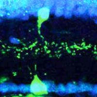
Object motion and amacrine cells in retina

Circuit in the Eye Relies on Built-In Delay to See Small Moving Objects

When we move our head, the whole visual world moves across our eyes. Yet we can still make out a bee buzzing by or a hawk flying overhead, thanks to unique cells in the eye called object motion sensors.
Grantee News

New clues found to vision loss in macular degeneration​​​

Scientists at Washington University School of Medicine in St. Louis have identified a pathway that leads to the formation of atypical blood vessels that can cause blindness in people with age-related macular degeneration.
A microglial cell (green) extends spider-like arms to capture and consume rod photoreceptor cells (blue). Credit: Dr. Wai Wong, NEI.

In Blinding Eye Disease, Trash-Collecting Cells go Awry, Accelerate Damage

Spider-like cells inside the brain, spinal cord and eye hunt for invaders, capturing and then devouring them. These cells, called microglia, often play a beneficial role by helping to clear trash and protect the central nervous system against infection.
Grantee News

Eye’s motion detection sensors identified

Researchers at Washington University School of Medicine in St. Louis have identified a neural circuit in the retina that enable the eye to detect movement.
Grantee News

Plugging In Your Vision's Autostabilization Feature

New research, published online May 7 in the journal Neuron, describes how axons of specialized nerve cells find their way through the brain’s maze of neurons to make the right connection.