
RPE or Retinal Pigmented Epithelium is a polarized monolayer of epithelia located in the back of the eye between the photoreceptors and the choroidal blood supply. RPE serves the crucial function of maintaining the health and function of the photoreceptors by maintaining the visual cycle, phagocytosis of photoreceptor outer segments and the chemical composition and volume of the sub-retinal space and choroid. RPE dysfunction leads to degenerative diseases of the eye including age-related macular degeneration
Technology: A method of developing functional RPE cells from induced pluripotent stem cells via in vitro differentiation protocols (iPSC-RPE cells)
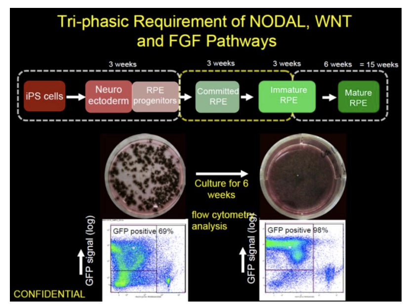
Competitive Advantage: Highly reproducible and efficient method (>95% pure) that generates iPSC-RPE that can be validated by genetic, structural and functional tests. iPSCRPE cells that can be used for in vitro studies or transplantation therapy (dry-AMD). iPSCRPE cell lines can be generated from fibroblasts or PBMC sources. iPSC-RPE reporter cell lines are also available for drug screening studies.
Intellectual Property: NEI/NIH has secured intellectual property of this technology which is available for co-development, non-exclusive/exclusive license consideration.
References
- The new paradigm: retinal pigment epithelium cells generated from embryonic or induced pluripotent stem cells. Bharti K, Miller SS, Arnheiter H.Pigment Cell Melanoma Res. 2011 Feb; 24(1):21-34.
- Developing cellular therapies for retinal degenerative diseases. Bharti K, Rao M, Hull SC, Stroncek D, Brooks BP, Feigal E, van Meurs JC, Huang CA, Miller SS. Invest Ophthalmol Vis Sci. 2014