About our work
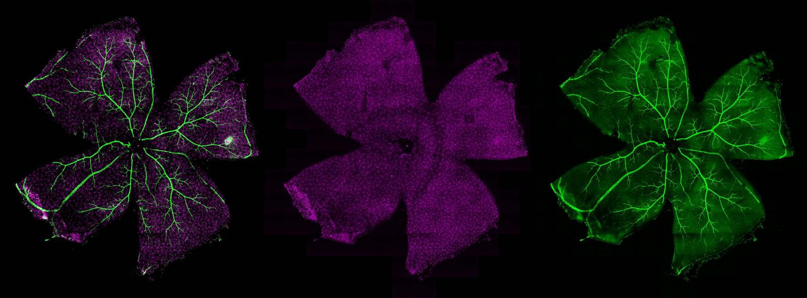
The National Eye Institute's Biological Imaging Core provides NEI scientists with a wide range of high resolution imaging and analysis applications including confocal microscopy, multi-photon imaging, Airyscan super-resolution, deconvolution, laser capture microdissection, in vivo and in vitro imaging. The primary objective of the Biological Imaging Core is to pair state-of-the-art instrumentation with novel imaging approaches to provide vision scientists new avenues for studying ocular disease processes.
Research Units
Biological Imaging Core Facility includes the following:
Biological Imaging Core Facility key staff
| Name | Title | Phone | |
|---|---|---|---|
| Robert Fariss, Ph.D. | Core Chief, Associate Researcher | farissr@nei.nih.gov | 301-496-2829 |
