Researchers at the University of North Texas Health Science Center are the first to characterize extracellular vesicles (EVs) in the tears of patients with keratoconus.

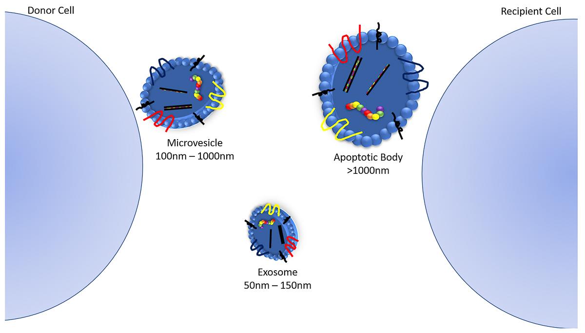
Illustration of extracellular vesicles, which are divided into three types: microvesicles, apoptotic vesicles, and exosomes. EVs are involved in cell-cell communication via transporting DNA, RNA, and proteins from one cell to another. Image credit: Brenna Hefley, University of North Texas.
Keratoconus occurs when the clear, dome-shaped front surface of the eye called the cornea bulges into a cone shape. The progressive, often painful condition can distort vision. The cause of keratoconus is unknown, but most researchers think it is a combination of genetics, hormonal imbalances and environmental factors, such as allergies and eye rubbing.
EVs are tiny membrane-bound messages that cells use to communicate. The study of EVs is exploding throughout many scientific disciplines around the world, as researchers are now beginning to explore their possible use in medical diagnostics and therapy.
Dimitrios Karamichos, executive director of the North Texas Eye Research Institute, and his team examined tears in the eyes of 10 healthy people (five males and females) and nine people with keratoconus (four males and five females). The team discovered that tear EVs have a distinct appearance, compared to their healthier counterparts. They also found that males have higher tear EV counts.
“I think this study is only the beginning of a massive, unexplored area of research in the context of keratoconus,” said Karamichos.
Tear EVs have been linked to other conditions, such as dry eye, Sjӧgren’s Syndrome and types of glaucoma.