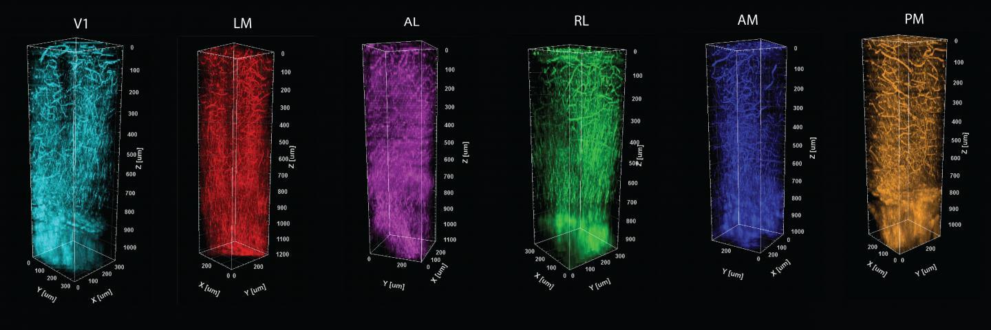
A distinct thicket of vessels and myelin fibers are evident in each of six color coded visual processing regions in the cortex of a mouse. The columns are formed by stacking images taken at 5-micron increments through a millimeter of depth in each of the regions. Credit: Sur Lab/MIT Picower Institute
To understand the massive capabilities and complexities of the brain, neuroscientists segment it into regions based on what they appear to do--like processing what we sense or how to move. What's been lacking, however, is an ability to tie those functional maps precisely and consistently to matching distinctions of physical structure, especially in live animals while they are performing the functions of interest. In a new study, funded by the National Eye Institute, MIT researchers demonstrate a new way to do that, providing an unprecedented pairing of functional mapping in live mice with distinguishing structural information for each region all the way through the cortex into deeper tissue below.
The study detailing the tools and techniques was published in Biomedical Optics Express.