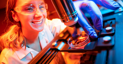
Madison Wilson, a Ph.D. student at UC San Diego, is first author of the study showing that human brain organoids implanted in mice have established functional connectivity to the animals’ cortex and responded to external sensory stimuli. Image credit: David Baillot/UC San Diego
A team of engineers and neuroscientists led by Duygu Kuzum from the University of California, San Diego, has demonstrated for the first time that human brain organoids implanted in mice have established functional connectivity to the animals’ cortex and responded to external sensory stimuli. The implanted organoids reacted to visual stimuli in the same way as surrounding tissues, an observation that researchers were able to make in real time over several months thanks to an innovative experimental setup that combines transparent graphene microelectrode arrays and two-photon imaging.
Human cortical organoids are derived from human induced pluripotent stem cells, which are usually derived themselves from skin cells. These brain organoids have recently emerged as promising models to study the development of the human brain, as well as a range of neurological conditions.
The UC San Diego-led team developed experiments that combine microelectrode arrays made from transparent graphene, and two-photon imaging, a microscopy technique that can image living tissue up to one millimeter in thickness.

Researchers were able to detect and image the border between a transplanted human brain organoid and mouse brain. Image credit: Madison Wilson/UC San Diego
By placing an array of these electrodes on top of organoids transplanted into mouse brains, researchers were able to record neural activity electrically from both the implanted organoid and the surrounding host cortex in real time. Using two-photon imaging, they also observed that mouse blood vessels grew into the organoid providing necessary nutrients and oxygen to the implant.
Their findings suggest that the organoids had established synaptic connections with surrounding cortex tissue three weeks after implantation, and received functional input from the mouse brain.